|
Tooth Anatomy - Tissues of a Tooth
The Anatomy of a Tooth
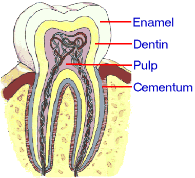
A basic knowledge of the internal anatomy of the tooth and its structure is necessary in order to understand the development of several dental conditions, their causes and their treatments. If we take a section of a human tooth we will be able to distinguish 4 different types of tissues, enamel, cementum, dentin, and pulp tissue. The main characteristics of each part of the tooth structure are described in this article (for more detailed information about each one follow the links to the specific pages). The main parts of tooth anatomy are Enamel, Dentin, Cementum, and Pulp. The Tooth EnamelEnamel is the outermost layer of the tooth structure of the crown, the part of the tooth that is above the gums. Dental enamel is the hardest and most mineralized substance in the human body. It looks like bone but it is much harder, denser, and its composition is 96% inorganic. Dental enamel offers a protective cover to the entire top of the tooth with its thickest point at the cusps of the tooth. It becomes thinner at the edges of the tooth closer to the gum line, up to the cement-enamel junction (CEJ) where it is substituted by the cementum that covers the root. It is slightly translucent, so any change in the color of the underlying dentin, either due to the development of tooth decay, or due to discoloration usually becomes visible through the enamel. Its natural color can vary from grayish white to yellowish. Although dental enamel is the hardest tissue of the human body, it is very vulnerable. Due to its very dense mineral structure, it is highly susceptible to acidic damage (demineralization), the main cause of tooth decay. Enamel is also very brittle, especially when not supported by sound underlying dentin. The main disadvantage of dental enamel is that it lacks the ability of repairing itself, as bones have. Any structural damage to the tooth enamel is permanent and can’t be repaired naturally by our body. The CementumCementum is the outer layer of the tooth structure for the root part of the tooth which lies under the gum line. It is a bonelike material, almost as hard as enamel, but with a less inorganic composition. Cementum is light yellowish in color, slightly lighter than dentin. It is not translucent as the enamel. The main function of cementum is to protect the roots and anchor the teeth in their sockets inside the jaw bone. A network of fibers made of connective tissue (known as periodontal ligament) connects the cementum with the alveolar bone and keep the tooth steady in its socket (for more details see ΄periodontium΄). It is more susceptible than enamel to tooth decay, especially due to the higher accumulation of bacterial plaque and dental calculus along the gum line, or underneath it (sub-gingival plaque). Unlike enamel, cementum is formed continuously throughout the life of the tooth to compensate for the substance loss due to tooth wear, and to allow for the attachment of new fibers of the periodontal ligament to the surface of the root. The DentinDentin is a bone-like material that makes up most of the tooth's structure. It is covered by the enamel in the crown and by the cementum in the root area of the tooth. It engulfs and protects the living part of the tooth, the pulp tissue. Dentin has a light yellow color, which gives the tooth its natural color prevailing through the almost translucent enamel. It consists of 70% inorganic materials, with a spongy, porous structure made of tubules extending from the enamel to the pulp chamber. Nerve endings enter the dentinal tubules and may transmit signals such as pain in response to external stimuli. Dentin is harder than bone but softer than enamel. It becomes very vulnerable to tooth decay if the protective cover of enamel or cementum is lost or weakened. However, the dentin is a living tissue, so it has the ability for some degree of growth and repair in response to certain physiologic and pathologic conditions. The Dental PulpThe dental pulp is the soft living tissue of the tooth. It can be found in the pulp chamber, a cavity in the center of the crown of each tooth, and inside the root canals which connect the pulp cavity with the opening at the root tip (apex) and through that with the rest of the body. The pulp contains blood vessels, connective tissue, nerves, and other cells including odontoblasts, fibroblasts, macrophages and lymphocytes. The main function of the pulp is providing sensation and nourishment to the tooth, and the formation of dentin and cementum. The pulp tissue responds to irritation either by forming reparative secondary dentin for protection against the source of irritation or by becoming inflamed. The pulp, as the living part of the tooth, has a high risk of infections. If bacteria manage to reach the pulp, e.g. due to tooth decay or through a crack of the tooth, they cause inflammation which is usually very painful due to the high pressure that is developed inside the enclosed space of the pulp chamber. If the pulp is infected and a dental abscess has developed, a root canal therapy is necessary to prevent further damage.
|
|
Dental Diseases |

 Although humans have 4 different
