|
Parts of the Mouth - Dental Anatomy
The main Parts of the Mouth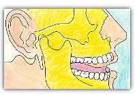
The main parts of the mouth anatomy include the following: The LipsThe lips are the soft parts of tissue at the front edge of the cheeks that form the anterior boundary of the mouth. They are covered externally by skin and internally by the same mucous membrane (mucosa) that lines the entire oral cavity. The lips help keep saliva and food inside the mouth and move it over the teeth for chewing. They are also important in the formation of speech and in several behavioral expressions such as kissing or laughing. The surface of lips in humans is equipped with very sensitive sensory receptors that help us determine temperature and texture of food, in order to avoid the ingestion of excessively hot or cold substances, or of rough, hard, or sharp objects. The inner surface of each lip is connected in the middle line to the corresponding gum by a fold of mucous membrane, the labial frenulum. The CheeksThe cheeks form the sidewalls of the mouth and cover the area of the face below the eyes, between the nose and ears, along the front of the face to the lips. They are composed of connective tissue, subcutaneous fat and certain muscles, with the outside layer covered by skin and the inside by a moist inner lining, a mucous membrane called ‘mucosa’. The cheek muscles are important in smiling, swallowing, compression and keeping food in the mouth for chewing and digestion. The TeethTeeth are the more characteristic part of the mouth's anatomy. They are white colored calcified organs embedded partially in sockets of the lower and upper jaws. The crown is the visible part of the tooth above the gums, while the root of the tooth keeps it in place with the help of the periodontal ligament that anchors it to the alveolar bone. Teeth in humans and other mammals are a critical component of the digestive system performing the function of tearing and chewing foods (mastication). They also contribute in speaking by helping in the formation of sounds. Properly aligned bright white teeth are a key asset in a person’s appearance. Finally, by supporting the lips and the cheeks, they play a role in the aesthetics of the face. The 20 primary (deciduous) teeth are the first to emerge; later they are replaced by the 32 permanent teeth of the adult human dentition between 6 to 13 years of age. There are 4 distinct types of adult teeth with different morphology and function: incisors, canines, premolars, and molars. Each tooth is composed of 4 types of dental tissues: enamel, dentin, cementum, and pulp. The PeriodontiumPeriodontium is a group of supporting tissues that surround teeth and keep them in place. The Gingiva - GumsThe gingiva, known as gums, is the pink soft tissue that surrounds teeth and covers the jaw bone. Gums are a delicate tissue that can easily get irritated, inflamed, and start to bleed if infected by the bacteria of dental plaque, developing a condition called gingivitis. When not treated, the condition can progress to periodontitis, a more severe form of gum disease, which leads to bone loss and possibly tooth loss. Red, swollen, bleeding gums that have lost their firm attachment with teeth are a clear sign of gum disease that needs immediate dental treatment. Careful, regular brushing and flossing are needed to avoid gum problems. The gums are a mucosal tissue that consists of collagen and elastin. They are attached to the cementum of the tooth and to the alveolar bone. Gingiva is part of periodontium; a group of tissues that support teeth in place. The Periodontal LigamentThe periodontal ligament is a layer of connective tissue fibers which hold the tooth anchored inside the alveolar socket of the jaw. One end of these fibers is attached to the cementum covering the root of the tooth, while the other end is attached to the alveolar bone around the root. The periodontal ligament works also as a protective elastic cushion for the tooth, allowing it to withstand the pressure of biting and chewing. The fibers of the periodontal ligament are the first ‘victim’ in case of periodontitis. The toxins produced by the bacteria of dental plaque destroy gradually the ligament creating periodontal pockets. Teeth become loose and in advanced cases they may fall off. Alveolar process and socketAlveolar sockets are the hollow areas of the jaw bones which host the roots of the teeth. Their bony walls that develop around the teeth to support them are called alveolar process. The TongueThe tongue is a thick solid muscular organ in the mouth. Its functions include chewing, digesting, swallowing, tasting and speaking (with most important its contribution in the digestive process). The tongue is attached to the back part of the floor of the mouth by a membrane called the lingual frenum, but its front tip can move freely allowing the tongue to be able to change size, shape, and position. At rest, it is positioned between the teeth and the lower jaw. It consists of voluntary muscle fibers and it is covered by a mucous membrane. The tongue is a very flexible muscle with large freedom of movement which allows it to move food around the mouth and place it between the chewing surfaces of teeth to help chewing. It mixes food with saliva, gathers the chewed food into a ball (bolus), and moves it towards the pharynx for swallowing. By adjusting the shape of the tongue and moving it in certain positions, we can form specific sounds and words that make up speech. The tongue is also a sensory organ, providing us with the sensation of taste. On the sides and upper surface of the tongue are a large number of ‘taste buds’ that can sense the 4 main tastes: sweet, sour, salty, and bitter. Finally, with the help of saliva, it assists in cleaning food residuals from teeth and the rest of the mouth. The JawsThe jaws are the two bony structures that form the mouth and carry the 32 human teeth. Lower Jaw (Mandible)The lower jaw also known as mandible is the largest and strongest bone of the face. The mandible in humans supports the 16 permanent lower teeth. The word ‘jaw’ or ‘jawbone’ used in the singular typically refers to the lower jaw. The lower jaw is a mobile component of the mouth. It is a U-shaped bone structure that stretches from one ear, down to the chin area and then back up again to the other ear. It is joined to the upper part of the head around the ear region by two jaw joints called ‘temporomandibular joints’ or TMJ. The ability of mandible for 3-dimensional movement is essential for our ability to chew properly. The human mandible can move forward to engage the incisors, up and down to crush and chew, and side-to-side to grind foods. Upper Jaw (Maxilla)The upper jaw, called ‘maxilla’, is fixed with the skull. The maxilla contains the alveolar process which holds the top row of teeth. The upper jaw consists of two large facial bones (maxillae) one on each side of the face which are fused together at the middle of the face with a suture. Incomplete closure of this suture is the cause of a malformation known as ‘cleft palate’. The maxilla forms part of the cheeks and roof of the mouth, part of the nasal cavity and the floor of the orbital cavity (where the eyeballs fit). Temporomandibular JointThe temporomandibular joints (TMJ) are a pair of movable joints between the mandible (lower jaw bone) and the temporal bone of the skull, one at each side of the face located at the area just in front of the ear. Problems associated with this joint can cause severe chronic pain (Temporomandibular Joint Dysfunction or TMJD). The PalateThe palate is the term used for the roof of the mouth, which separates it from the nasal cavity. It is divided into two parts: the hard palate and the soft palate.
The Salivary GlandsThe salivary glands provide our mouth with saliva. There are three pairs of major salivary glands (one on each side of the face):
The SalivaSaliva is a biological fluid secreted in the mouth by the salivary glands. Saliva is almost 99,5% water, but it also contains some very important proteins, antibodies, and enzymes. Lysozyme is one of these enzymes that provide saliva with antimicrobial action against infections. Saliva contains amylase which helps in the digestive process by breaking down polysaccharides (starch) into short chains of glucose. Other main functions of saliva are:
The Floor of the MouthThe floor of the mouth is formed by the tongue that covers most of it and mucous membranes that extend from the base of the tongue to the gums of the lower jawbone. Muscles of the mouthNone of the functions of the mouth could be performed without the help of the mouth muscles. All the perfectly controlled movements of the various parts of the mouth (jaw, lips, tongue, cheeks) so that we may eat and speak are made possible by the many muscles which lie under the mucous membrane of the mouth. The movements of the mandible are mainly produced by the 4 muscles of mastication: the masseter, temporalis, lateral pterygoid, and medial pterygoid muscles.
|
|

|
Dental Diseases |

|